From molecular diagnosis to microsurgical reconstruction: keys to treating sarcoma
Comprehensive care is key to treating these rare tumours.
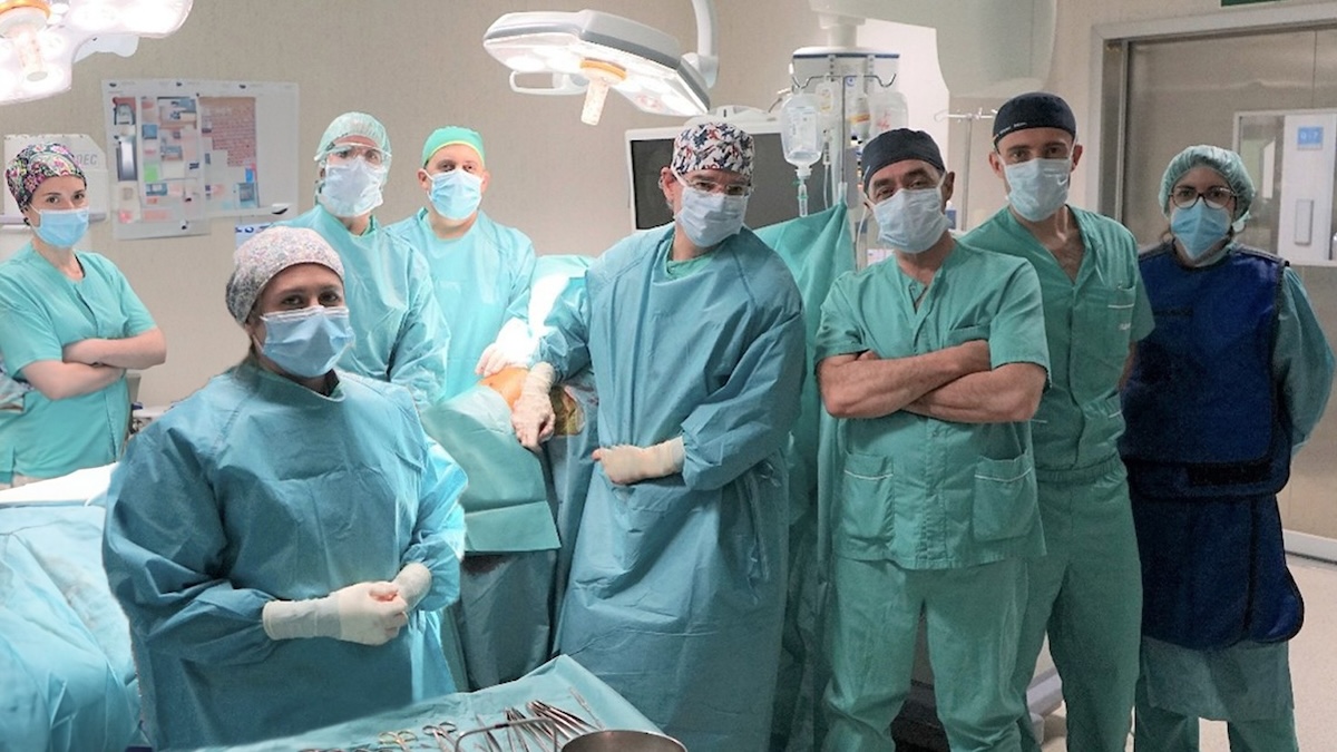
Sarcomas account for around 1% of cancer cases. To treat them properly, a multidisciplinary approach is needed, involving different areas such as medical and radiation oncology, orthopaedic oncology surgery, vascular surgery and plastic surgery, radiodiagnostics and pathological anatomy, among others, according to various specialists at Ruber Internacional Hospital on the occasion of National Sarcoma Day, which is commemorated on Saturday, 20 September. This commemoration, promoted by the Mari Paz Jiménez Casado Foundation with the aim of raising awareness of the disease and sensitising society, involves the participation of 40 centres in the 17 autonomous communities, which are joining in the day with informative and awareness-raising activities.
Precision diagnosis from the first biopsy
Dr Purificación Domínguez, head of the Pathological Anatomy Service, emphasises that "pathological anatomy plays an essential role in classifying sarcomas and distinguishing them from other lesions. The first diagnostic step is to obtain a sample of the tumour through biopsy, which we analyse using histological techniques to identify the cell type, growth pattern and structural characteristics. These characteristics, together with clinical and radiological data, allow us to differentiate between the multiple subtypes of sarcomas and distinguish them from benign lesions or pseudosarcomas."
Regarding diagnostic difficulties, the specialist adds that the main difficulty is their rarity, which means that these cases must be concentrated in centres with experience. ‘In addition, some benign lesions can mimic sarcomas, and there are tumours of intermediate malignancy that require a very precise approach.’
In terms of technological advances, the pathologist points out that "immunohistochemistry and molecular techniques such as FISH, PCR, or NGS are now essential. The detection of specific genetic alterations, such as EWS-FLI1 fusion in Ewing's sarcoma or MDM2 amplification in dedifferentiated liposarcoma, is key to diagnosis and molecular classification. All of this opens the door to true precision medicine, which is essential for planning the most appropriate treatment."
Preserving organs and rebuilding functionality
Dr. César Casado, head of Plastic Surgery at the hospital, highlights the role of soft tissue reconstruction after tumour resection. ‘We use robust flaps that can withstand subsequent radiotherapy, often free flaps with microsurgical techniques. The priority is to ensure that the oncological resection is complete, and from there we restore the functionality and aesthetics of the affected area.’
As he explains, ‘limb preservation techniques have brought about a radical change: today, amputation is the exception, not the norm. In addition, we use nerve transfers and sutures to reinnervate muscles, and lymphatic recovery techniques that prevent sequelae. All of this results in a better quality of life for the patient.’
Complex resections and reconstructions
Dr Pablo Gallo, head of the Angiology and Vascular Surgery Service, points out that ‘the resection of sarcomas with vascular involvement poses challenges such as the risk of bleeding, the difficulty of differentiating between compression and infiltration, and the need to balance oncological radicality with functional preservation. Sometimes, the true vascular involvement is only confirmed in the operating theatre.’
For these cases, he points out, "vascular reconstructions are essential. If a major artery is compromised, we perform bypasses or other revascularisation techniques; and when it comes to major veins, we add medical treatment, thrombotic prophylaxis and even vena cava filters. All of this requires coordination with orthopaedic oncology surgery to plan the resection and reconstruction in a single surgical procedure. Thanks to this collaboration, we can preserve limbs and avoid unnecessary amputations," he says.
Orthopaedic oncological surgery: teamwork that makes a difference
Dr. Eduardo Ortiz, head of Orthopaedic Oncological Surgery at Ruber Internacional, emphasises the importance of treating sarcoma patients in multidisciplinary units.
‘Sarcoma is a rare and complex tumour. A coordinated team reduces time, anxiety and improves survival. Care is rigorously structured thanks to coordination between specialities and the support of nursing and administration, who accompany the patient throughout the process.’
In terms of technological advances, 3D navigation-guided surgery, customised cutting guides and three-dimensional models stand out, allowing for safer and more precise resections. In paediatrics, expandable endoprostheses preserve growth and biological grafts remain key.
The Unit also draws on the expertise of Medical Oncology, led by Dr María Angeles Vaz, who recognises that the treatment of sarcomas requires a highly specialised and personalised approach. Thanks to our in-depth knowledge of tumour biology, we now know that there is no single type of sarcoma, but more than 70 different subtypes, each with its own molecular mechanisms. ‘This diagnostic precision allows patients to be classified into three main groups: those who benefit from precision targeted therapies against specific molecular alterations; those who may respond to immunotherapy; and those for whom chemotherapy remains the main option, albeit with more advanced strategies, such as its use before surgery to facilitate more effective and conservative interventions,’ she explains.
The oncologist emphasises the importance of coordination between medical oncologists, surgeons, radiotherapists, radiologists and anatomopathologists, which is coordinated through the Multidisciplinary Tumour Committee, where all cases are analysed jointly from the moment of diagnosis. ‘No decision is taken in isolation, which guarantees the most appropriate and personalised planning for each patient,’ she says.
Likewise, in Radiation Oncology, Dr Belén Belinchón affirms that the great advantages offered by state-of-the-art radiotherapy techniques for patients with sarcomas are greater precision in tumour irradiation and greater protection of healthy tissue near the tumour. Radiotherapy with intensity-modulated techniques, image guidance and motion control achieves more effective treatment, with better oncological results and fewer side effects.
"Radiotherapy treatment is necessary as a complement to surgery in a large number of patients with sarcoma. Radiotherapy can be administered before or after surgery, and in some cases, exclusively. In addition, in patients with recurrent sarcoma, re-irradiation with advanced techniques can now be considered. Also, when there is a limited number of metastases, robotic radiosurgery with Cyberknife allows these lesions to be treated safely, accurately and precisely in just a few days."
Dr Fernando Herráiz, from the Radiodiagnostics Department, explains the role of medical imaging in the entire sarcoma care process: from the detection and accurate characterisation of the lesion to treatment planning and follow-up. To do this, advanced techniques such as magnetic resonance imaging, tomography, ultrasound or PET are used, and in some cases, image-guided biopsies that allow the diagnosis to be refined and the most appropriate treatment to be defined.
‘The aim of the specialists is to identify and characterise the lesion as accurately as possible,’ explains Dr Herráiz. ‘In certain cases, we even resort to obtaining image-guided tissue samples, which allows us to establish a key clinical-radiological correlation to confirm the diagnosis and define the most appropriate therapeutic strategy.’
Looking to the future
The multidisciplinary nature of the unit allows all clinical decisions to be integrated into joint committees, ensuring strategies tailored to each case and constant support for patients and families.
The next goals are ambitious: to consolidate limb-sparing surgery, implement new technologies such as augmented reality and 3D planning, promote clinical research and train new specialists.
‘Through technological innovation and multidisciplinary collaboration, we can ensure that patients have access to the best possible therapeutic options,’ concludes the team at the Sarcoma Unit at Ruber Internacional Hospital.