Dynamic Digital Radiography (DDR)
Dynamic digital radiography is an innovative imaging diagnostic technique that allows real-time moving X-rays.
Dynamic digital radiography, or DDR (Dynamic Digital Radiography), uses X-rays to capture moving images of the body’s interior. This effect is achieved because the radiological equipment emits several rapid, successive pulses of energy for a few seconds. When all the obtained radiographs are combined, the movement of the body’s structures is perceived in real time.
A conventional radiograph provides morphological information of the body, while DDR additionally provides quantified functional data. The amount of radiation the patient receives is similar to that of a conventional X-ray and significantly lower than that of fluoroscopy, which emits X-rays continuously and requires contrast material to clearly distinguish soft tissues.
The precise images of dynamic digital radiography reduce the time and facilitate a personalized diagnosis, as highly accurate information is available.
How does it work?
Just like conventional X-rays, a device is used to emit X-rays and a radiation-sensitive plate captures the images after passing through the body tissue. The patient can remain lying down or standing, which is a clear advantage over magnetic resonance imaging or computed tomography. The images are digital, so they are processed and displayed on a computer screen.
The main innovation of dynamic digital radiography is that, during 15 seconds, it emits 15 pulses of radiation per second. When these 225 slides are viewed consecutively in a cine loop, a moving X-ray is obtained.
What are the benefits of dynamic digital radiography?
The most notable benefits of DDR are:
- Moving images that provide information about the anatomy and functionality of body structures.
- Wide field of view.
- Large radiographs (up to 17 x 17 inches or 43.8 centimeters).
- High resolution.
- Low radiation dose.
- Short examination time (around one minute to capture about 20 seconds of movement).
- The patient can be lying down, sitting, or standing.
When is it indicated?
Dynamic digital radiography is primarily used in the following specialties:
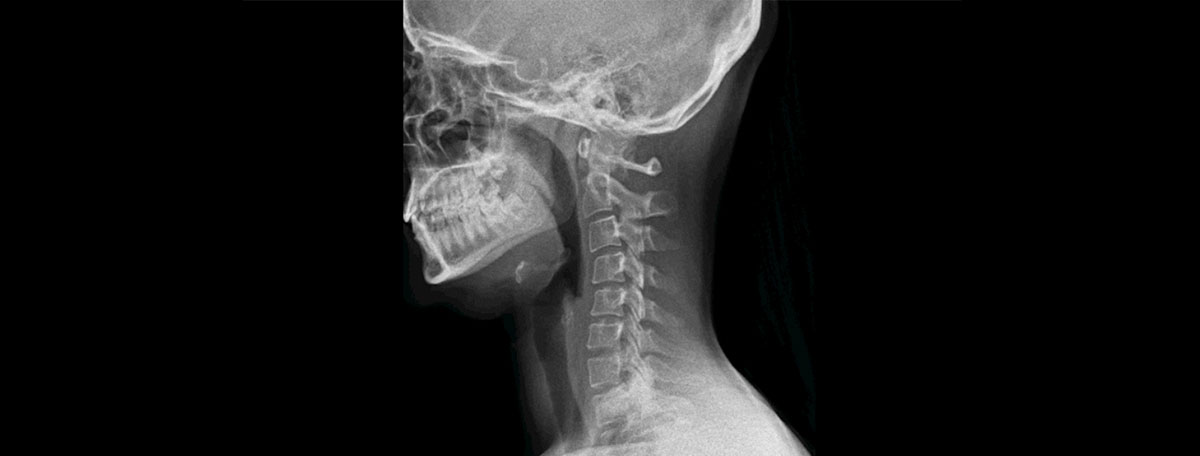
- Orthopedics: to study the wrist, shoulder, or knee joints, as well as spinal pathologies such as whiplash, herniated discs, or scoliosis.
- Pulmonology and intensive care medicine: to evaluate lung function, as two or three complete breaths can be observed in a single exam. It is useful for diagnosing dyspnea, pulmonary hypertension, or chronic obstructive pulmonary disease.
What to expect from dynamic digital radiography
When undergoing DDR, the patient does not notice any differences compared to conventional X-rays. The test is outpatient, and routine activities can be resumed once it is completed.
To access the radiology room, the patient must wear an appropriate gown and remove any metallic objects (jewelry, glasses, dentures). Once positioned in the best way to take the images, the patient must follow the movement instructions provided by the healthcare staff from the adjacent room. After all images are taken, one or two minutes later, the X-ray emitting device and the receptor plate are removed, and the patient can leave the medical center.
Specialties in which it is used
Radiologists are responsible for taking the images, usually at the request of specialists in emergency medicine, intensive care, pulmonology, orthopedics, or thoracic surgery.