



A minimally invasive technique makes it possible to destroy a pancreatic tumor without surgery
Hospital Ruber Internacional has performed its first endoscopic ultrasound-guided radiofrequency ablation to treat an insulinoma, a rare pancreatic neuroendocrine tumor that can cause episodes of hypoglycemia.
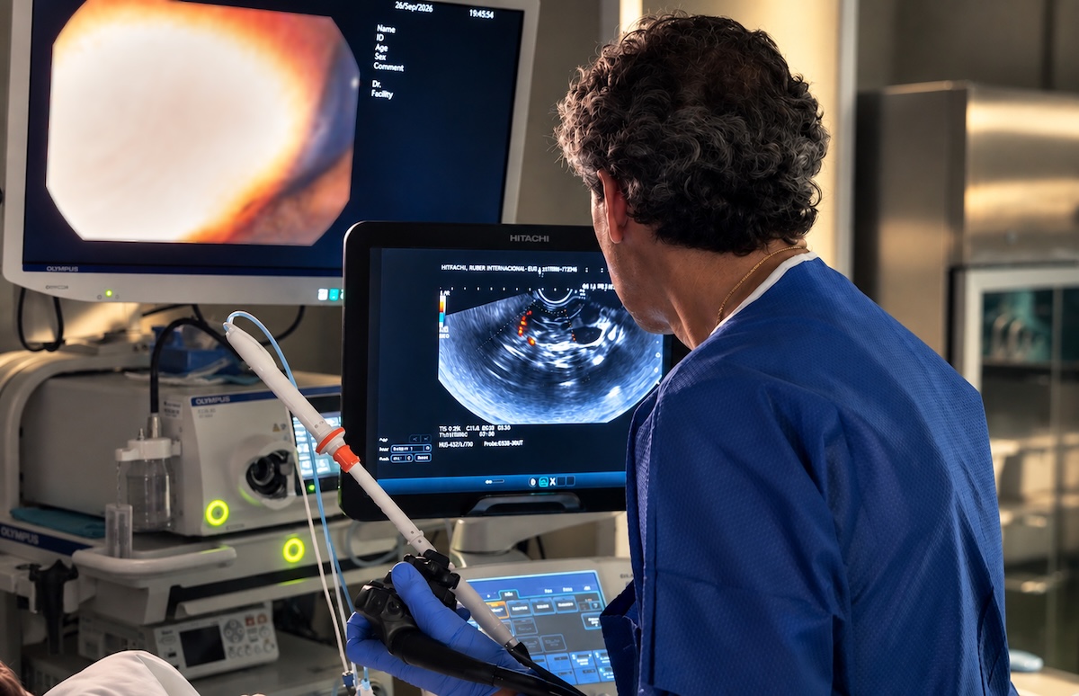
An apparently unexplained drop in blood sugar levels may, in rare cases, be caused by a small tumor located in the pancreas. This is what happens with insulinomas, neuroendocrine tumors that produce excessive and inappropriate amounts of insulin, potentially leading to recurrent episodes of hypoglycemia.
Hospital Ruber Internacional has introduced a minimally invasive alternative for selected patients with this type of lesion. For the first time, the hospital has performed an endoscopic ultrasound-guided radiofrequency ablation of a pancreatic neuroendocrine tumor, a procedure that allows specialists to reach the lesion through the stomach or duodenum and destroy tumor cells using thermal energy, without the need for pancreatic surgery.
 High resolution image. This link will open using lightbox, there may be a context switchThe procedure was performed by Dr. José Miguel Esteban, Head of the Endoscopy Unit at Hospital Ruber Internacional, after Dr. Juan Carlos Percovich and Dr. Helena Urriza, endocrinologists at the hospital, identified the condition and initiated a multidisciplinary diagnostic and therapeutic approach.
"We access the pancreas through the stomach or duodenum and, using the ultrasound imaging provided by endoscopic ultrasound, insert a needle directly into the tumor. This needle is connected to an energy source that allows us to destroy the tumor cells in a controlled manner," explains Dr. Esteban.
Hypoglycemia Without an Apparent Cause
An insulinoma is a rare pancreatic neuroendocrine tumor. Although these lesions are usually small, their ability to release insulin can cause blood glucose levels to fall abnormally.
The patient treated at Hospital Ruber Internacional had recently begun experiencing episodes of hypoglycemia with no known cause. The diagnostic investigation identified an insulin-producing lesion in the pancreas responsible for these drops in blood glucose.
"When episodes of hypoglycemia occur without an obvious explanation, it is essential to investigate their underlying cause. In this patient, the endocrinological assessment raised suspicion of inappropriate insulin production and enabled us to quickly focus the diagnostic tests on locating the responsible lesion," explain Dr. Juan Carlos Percovich, Head of the Endocrinology and Nutrition Department, and Dr. Helena Urriza.
Early detection proved crucial in accelerating the entire process. From the moment the patient was admitted, the specialists coordinated the necessary diagnostic tests.
 High resolution image. This link will open using lightbox, there may be a context switchAn MRI scan was performed on the same day, followed the next day by an endoscopic ultrasound with fine-needle sampling of the lesion, which made it possible to obtain tissue samples and confirm the diagnosis of a neuroendocrine tumor.
"Within just a few days, we were able to move from investigating hypoglycemia of unknown origin to locating the tumor, obtaining diagnostic confirmation, and discussing, as a multidisciplinary team, the most appropriate therapeutic option for the patient," emphasizes Dr. Juan Carlos Percovich.
The case was subsequently presented to the gastrointestinal tumor board, where the specialists involved jointly assessed the available treatment options and decided to proceed with radiofrequency ablation.
Reaching the Pancreas Through the Digestive Tract
Endoscopic ultrasound is a technique that combines gastrointestinal endoscopy with ultrasound imaging. It allows specialists to visualize structures located close to the stomach or duodenum, such as the pancreas, and identify even very small lesions.
This proximity also makes it possible to introduce needles under imaging guidance to obtain tissue samples or, as in this case, deliver treatment directly to the tumor.
"Endoscopic ultrasound provides us with a highly precise image of the lesion and allows us to reach it with a needle through the stomach or duodenum. Instead of using the needle solely to obtain a sample, we can also use it to deliver radiofrequency energy directly to the tumor," says Dr. José Miguel Esteban.
Once the needle is positioned inside the lesion, the energy produces a controlled increase in temperature, resulting in the destruction of the targeted tumor tissue. The entire procedure is performed under endoscopic ultrasound guidance.
Avoiding Pancreatic Surgery in Selected Patients
Until now, surgery has played a central role in the treatment of insulinomas. However, because these lesions are located in the pancreas, surgical intervention can be associated with significant morbidity, a considerable hospital stay, and a prolonged recovery period.
Radiofrequency ablation offers an alternative for carefully selected patients, following an individualized and multidisciplinary assessment.
"This is a procedure performed under sedation that does not require surgery. If we achieve a good response, we can spare the patient the risks associated with pancreatic surgery and significantly reduce recovery time," explains the Head of the Endoscopy Unit.
Following the procedure, the patient is initially monitored to assess their recovery. In these cases, provided no complications arise, the hospital stay may be limited to a single night.
For Dr. Esteban, introducing this technique does not mean replacing surgery as the standard treatment in all cases, but rather expanding the therapeutic options available to certain patients.
"Not all pancreatic neuroendocrine tumors, nor all patients, are suitable candidates for radiofrequency ablation. The key is to carefully select each case, evaluate the different alternatives, and jointly determine which treatment offers the best balance between effectiveness and safety," he emphasizes.
Blood Glucose Levels: One of the First Indicators of Treatment Response
Insulinomas have a particular characteristic that helps specialists assess the patient's progress after treatment: their direct relationship with blood glucose levels.
Following ablation, specialists closely monitor blood glucose, which can now be done conveniently using continuous glucose monitoring sensors. Follow-up also includes laboratory measurements of insulin and other related parameters.
"If the tumor was responsible for producing insulin inappropriately, one of the first things we monitor after treatment is precisely how blood glucose levels behave and whether the episodes of hypoglycemia disappear," explains Dr. Esteban.
However, follow-up does not end when symptoms resolve. Patients require ongoing clinical and imaging surveillance to assess the evolution of the lesion. Therefore, in addition to laboratory tests, subsequent imaging studies such as MRI and endoscopic ultrasound are recommended.
The introduction of endoscopic ultrasound-guided radiofrequency ablation enables Hospital Ruber Internacional to expand its treatment options for selected pancreatic neuroendocrine tumors through a less invasive approach.
"Our goal is to have different therapeutic tools available so that we can choose the most appropriate one for each patient. In selected cases, this technique allows us to treat the tumor directly through a minimally invasive approach and avoid pancreatic surgery, always as part of a shared decision made by the different specialties involved," concludes Dr. Esteban.