


Recognizing Emotions During Brain Surgery to Preserve the Brain’s Most Complex Functions
Dr. Jesús Martín-Fernández, neurosurgeon at Ruber Internacional Hospital and President of the World Society of Awake Brain Mapping, applies an innovative three-step multimodal cognitive mapping model that adapts surgery in real time according to the patient’s brain responses.
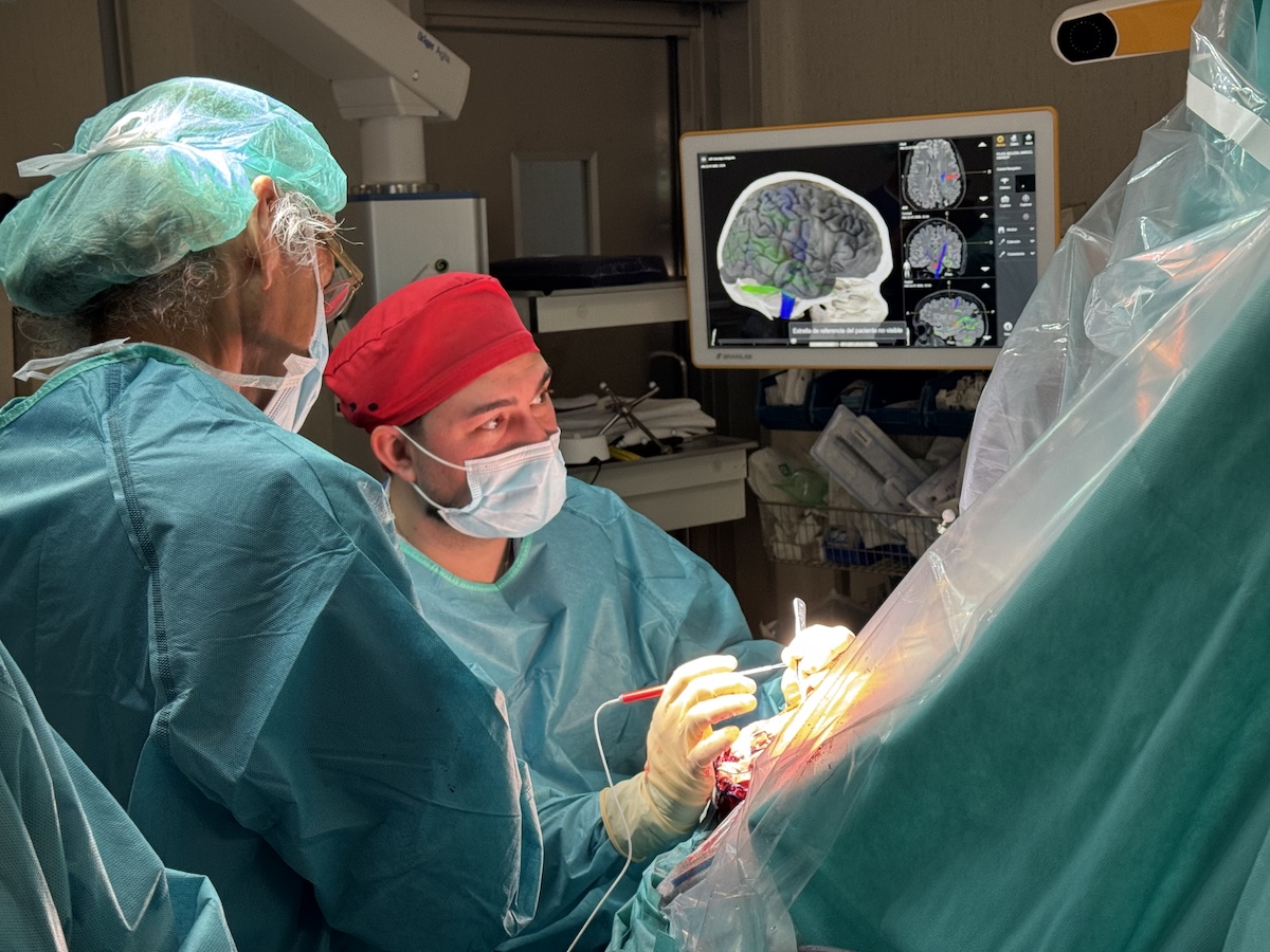
Marcel, a 32-year-old patient, looks at a series of faces displayed on a tablet and identifies the emotions they express: "uncertain," "embarrassed." As he answers, he remains awake in the operating room at Ruber Internacional Hospital during a critical stage of surgery to remove a brain tumor located in the right precuneus, one of the most complex regions of the human brain. His responses allow neurosurgeon Dr. Jesús Martín-Fernández to identify, in real time, the brain networks that must be preserved to maintain functions such as social cognition, autobiographical memory, self-referential processing, and emotion recognition.
The procedure is an example of the innovative three-step multimodal cognitive mapping model developed by Dr. Martín-Fernández, which transforms awake brain surgery into a dynamic procedure that continuously adapts to the brain's functional responses. Beyond preserving speech or movement, this technique makes it possible to protect higher cognitive functions that are essential for patients to maintain their independence, personal relationships, and quality of life.
Beyond Language: Assessing Emotions During Brain Surgery
During the operation, Marcel actively collaborated by performing different cognitive tasks while the surgical team stimulated various brain regions with low-intensity electrical current. In addition to moving his arm, counting aloud, and completing neuropsychological tests, he participated in an emotion recognition task specifically designed for the operating room. Whenever brain stimulation disrupted his ability to recognize emotions or triggered an emotional response—such as temporary episodes of crying—the team identified that they had reached a critical functional network and modified the surgical strategy to preserve it.
"What the patient tells us and how they respond to the tasks guides us in determining how far we can safely proceed," explains Dr. Jesús Martín-Fernández.
"Our goal is no longer simply to ensure that patients can speak or move a hand after surgery. We want to preserve the abilities that allow them to recognize people, understand others' emotions, maintain relationships, and continue with their life projects," he adds.
Until only a few years ago, these higher-order cognitive functions could not be systematically evaluated during brain surgery.
A Brain Mapping Approach That Evolves with the Brain
The three-step multimodal cognitive mapping technique represents a significant advancement over traditional awake brain surgery because it views the brain as a dynamic network rather than a collection of isolated functional areas.
According to Dr. Martín-Fernández, the procedure consists of three stages:
- Identification of critical nodes within brain networks using direct electrical stimulation.
- Continuous assessment of multiple cognitive functions throughout tumor resection using tasks tailored to each patient.
- Identification of connectome stop points—the deep functional boundaries that indicate when tumor removal should stop to avoid disconnecting essential neural networks.
Unlike conventional approaches, this methodology enables continuous reassessment of brain function throughout the procedure, allowing surgeons to adapt their strategy as the brain's functional organization changes during tumor resection.
"The brain changes functionally while we operate. It is not enough to create a functional map at the beginning of surgery; we need to update it continuously. Our model turns brain mapping into a dynamic process that evolves together with the brain," says the specialist.
Personalized Brain Surgery
 High resolution image. This link will open using lightbox, there may be a context switchEvery procedure is individually designed for each patient. The cognitive tasks performed during surgery vary depending on the tumor's location, the brain networks involved, and the functions that are most important to preserve for that individual, whether language, memory, attention, executive functions, or emotion recognition.
This personalized approach allows surgeons to maximize tumor removal while preserving each patient's unique functional profile.
"Every brain is organized differently, and every patient has different priorities. For some, preserving language is essential; for others, it may be recognizing the emotions of loved ones or maintaining the cognitive abilities needed to continue their profession. That is what truly personalized medicine means in neurosurgery," concludes Dr. Jesús Martín-Fernández.
Marcel's operation was not the only procedure performed that day using this personalized surgical model. During the same operating session, the team also treated a 60-year-old woman who had been monitored for more than a decade for a tumor located in a brain region critical for language. Surgery had been postponed for years because of the high risk of permanent functional deficits.
Thanks to the three-step multimodal cognitive mapping approach, the team was able to continuously monitor not only language but also other cognitive functions potentially affected by the tumor's location and connectivity. This enabled them to adapt the surgery in real time and maximize tumor resection while preserving the patient's functional abilities.
With these two procedures, Ruber Internacional Hospital further consolidates a new generation of personalized brain surgery based on network neuroscience, intraoperative cognitive monitoring, and the preservation of higher cognitive functions that, until recently, could not be systematically assessed during brain surgery.