


Brain Magnetic Resonance Imaging (MRI)
A brain MRI is a non-invasive diagnostic test that uses radiofrequency waves and a magnetic field to produce images of brain tissue and nerves.
General Description
Brain magnetic resonance imaging (MRI) is a non-invasive test that provides images of the brain, brainstem, and surrounding tissues. This test does not use ionizing radiation and is therefore safe for the body.
Images are obtained through a combination of a magnetic field and radiofrequency waves, which cause movement in the body’s hydrogen protons. A computer translates the resulting energy changes into cross-sectional images which, when combined, provide a three-dimensional view of the brain.
There are two types of brain MRI depending on the equipment used:
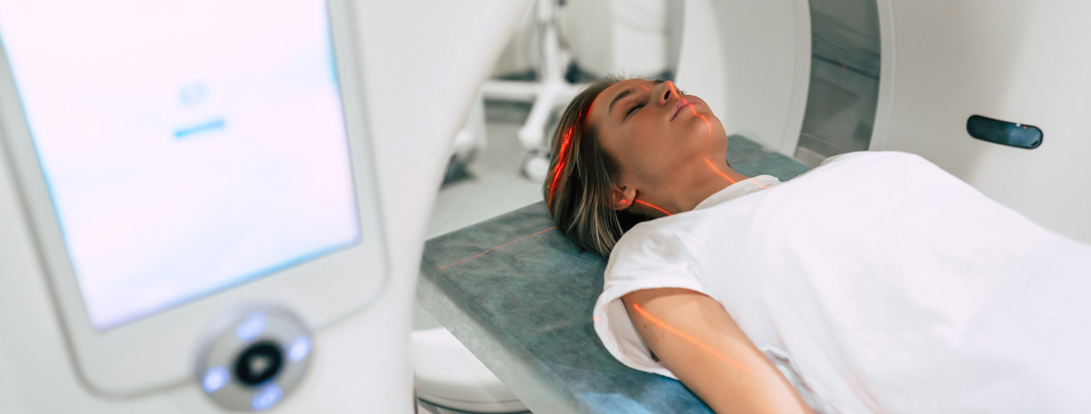
- Closed MRI: This involves a tube-shaped device approximately 70 centimeters in diameter into which the patient is inserted while lying on a movable table. This is the most effective type, as it allows the generation of a high-intensity magnetic field.
- Open MRI: This consists of two plates facing each other, separated by about 180 centimeters. It is used for patients with claustrophobia, but image quality tends to be lower due to the difficulty in generating a strong magnetic field.
Brain MRI is primarily a diagnostic procedure, although it is also used to assess treatment response in certain conditions.
When is it indicated?
Brain MRI is indicated for detailed examination of brain and nerve tissues. It is commonly used to identify the causes of:
- Recurrent headaches
- Seizures
- Dementia
- Muscle weakness, tingling, or numbness
- Visual disturbances
- Speech difficulties
- Hearing loss
- Behavioral changes
The analysis of brain MRI images helps detect conditions such as:
- Cysts and tumors
- Aneurysms (dilation of a blood vessel wall)
- Bleeding or hematoma formation in brain tissue, subarachnoid, subdural, or epidural spaces
- Infections
- Congenital abnormalities
- Stroke
- Multiple sclerosis
- Hydrocephalus (accumulation of fluid in the ventricles)
- Traumatic brain injury
- Age-related changes such as brain volume loss
How is it performed?
Brain MRI is performed with the patient lying on a table that is inserted into the MRI scanner. The device generates a magnetic field that aligns the body’s protons. When radiofrequency waves are emitted, the protons attempt to resist the magnetic force. As the waves stop, protons return to their original alignment, releasing energy at different rates depending on the type of molecule. This energy is used to create images with varying intensities for each tissue type.
In some cases, contrast-enhanced MRI is performed to obtain more detailed images. The contrast agent, usually gadolinium-based, is absorbed more readily by certain tissues—such as cancerous tissue—allowing them to appear more clearly.
Risks
Brain MRI is generally a safe procedure. However, it is contraindicated in patients with certain metal implants, such as pacemakers or prosthetic devices.
The contrast agent may cause mild allergic reactions such as itching, warmth, or nausea, although such reactions are uncommon.
What to expect from a brain MRI
Before the scan, patients must sign an informed consent form. They are provided with a hospital gown and must remove all metal objects. Makeup should be removed, as some products contain metallic components.
While lying on the table, a peripheral intravenous line may be placed for contrast administration if needed. Earplugs are provided to reduce the noise generated by the MRI machine as it rotates to produce the magnetic field. Despite this, loud tapping or knocking sounds are common during the scan.
The medical staff leaves the room, but the patient is monitored at all times via cameras. A microphone inside the scanner allows for communication in case of any issues. To ensure clear imaging, the patient must remain as still as possible throughout the 30 to 60-minute procedure. For this reason, sedation or anesthesia is often used in pediatric patients.
Medical specialties that request brain MRI
Brain MRI is typically requested by specialists in oncology, neurology, or pediatric neurology, and the procedure is performed by radiologists.
How to prepare
Patients are advised to wear comfortable clothing and avoid metal items or makeup, as some cosmetics contain metallic particles.
Fasting is only required if sedation is planned—typically for patients unable to remain still throughout the procedure.


